

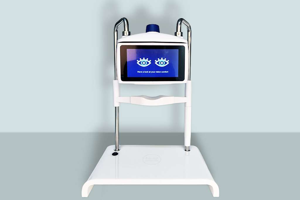
Dry Eye Screening
tearcheck®
Designed to set a new standard in dry eye examination, featuring two patented tests: TFSE® and OSIE®.
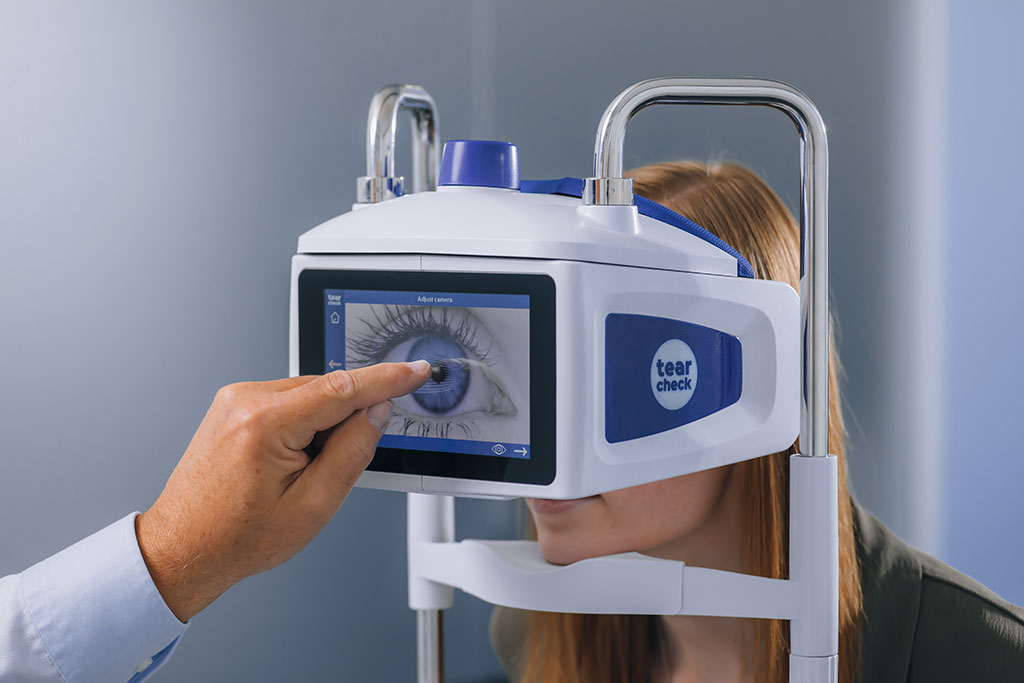
A visionary technology
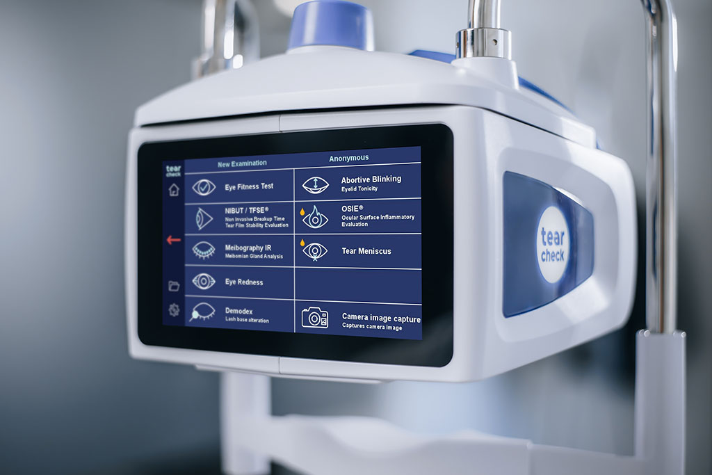
A complete range of precision exams at your fingertips
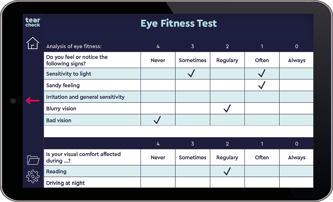
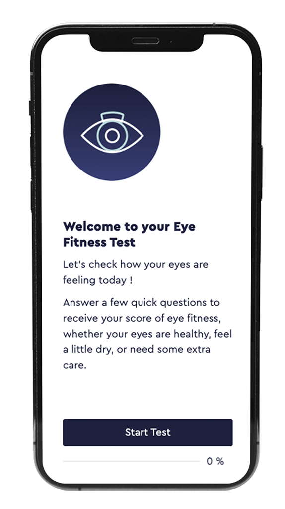
Provides an overview of the patient’s eye fitness in everyday life.

Share the same imaging sequence
Shows the micro-movements of the tear film
Quick: Evaluation over 10 seconds
Gives the user an exact score of dryness

Makes it possible to visualize the Meibomian Glands.
Shows the rate of gland loss (in %).
Shows the morphology of the glands present.

Makes it possible to assess hyperemia caused by dry eye induced inflammatory processes.
Re-assess the patients as the treatment progresses.
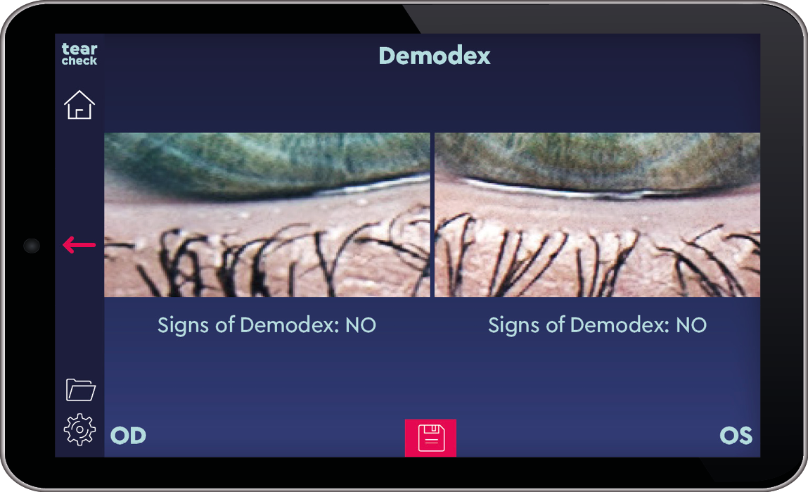
Enlarged image capturing the base of the eyelashes.
Makes it possible to trace and visualize the signs of demodex presence.
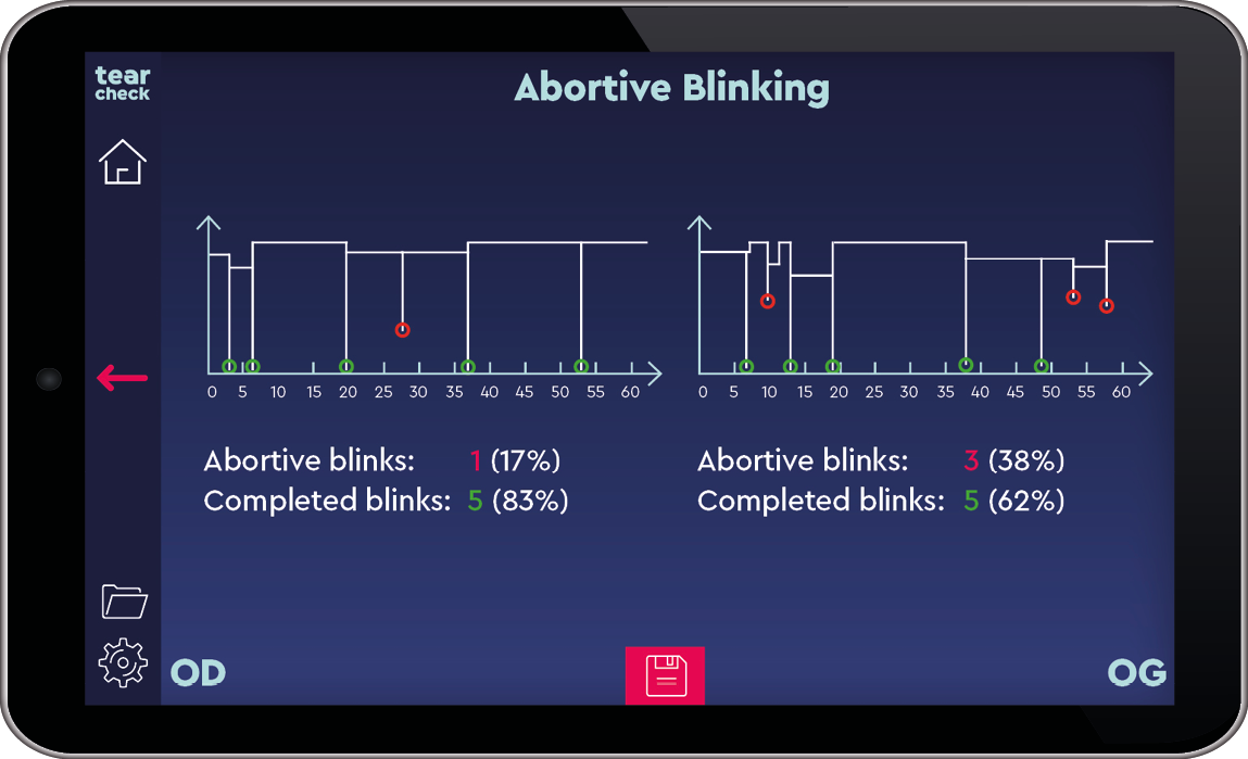
Shows all blinks identified over the acquisition time (expected 1 minute).
Determines the structure of the identified blinks: complete or incomplete blinks.

Shows increased risk of inflammation
Exact dimension and score of the dryness
The new standard tool for eye surgeons and lense-opticians
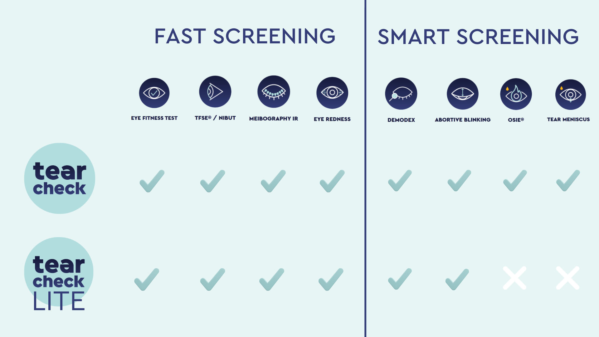
* not included in the tearcheck® LITE variant
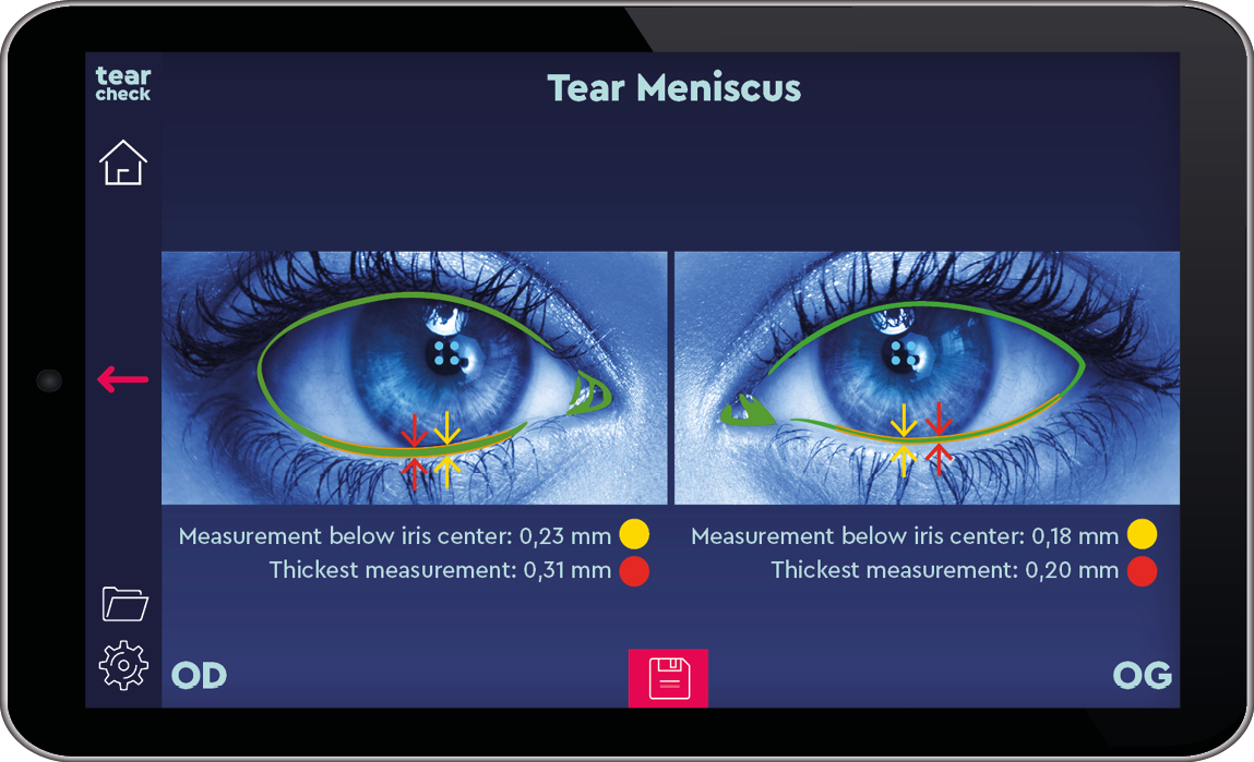
Shows the height of the tear meniscus.
Two values are calculated: measurement below the iris center and thickest measurement.
* not included in the tearcheck® LITE variant
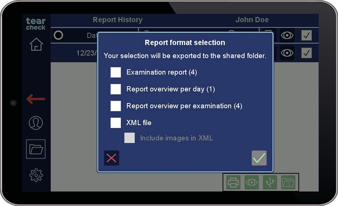
Allows you to select the report tupe that fits best your data management needs :
export by patient, by day or by exam.
Provides an overview of the patient’s eye fitness in everyday life.
Share the same imaging sequence. Shows the micro-movements of the tear film. Quick: Evaluation over 10 seconds. Gives the user an exact score of dryness.
Makes it possible to visualize the Meibomian Glands. Shows the rate of gland loss (in %). Shows the morphology of the glands present.
Makes it possible to assess hyperemia caused by dry eye induced inflammatory processes. Re-assess the patients as the treatment progresses.
Enlarged image capturing the base of the eyelashes. Makes it possible to trace and visualize the signs of demodex presence.
Blinking
Shows all blinks identified over the acquisition time (expected 1 minute).
Determines the structure of the identified blinks: complete or incomplete blinks.
Shows increased risk of inflammation
Exact dimension and score of the dryness
The new standard tool for eye surgeons and lense-opticians
* not included in the tearcheck® LITE variant
Shows the height of the tear meniscus.
Two values are calculated: measurement below the iris center and thickest measurement.
* not included in the tearcheck® LITE variant
Allows you to select the report tupe that fits best your data management needs :
export by patient, by day or by exam.
Discover tearcheck®
8 exams for smart analyses under 10 minutes
EYE FITNESS TEST
TFSE® / NIBUT
Tear Film Stability Evaluation / Non-Invasive Breakup Tim
MEIBOGRAPHY IR
EYE REDNESS
DEMODEX
ABORTIVE BLINKING
OSIE®
Ocular Surface Inflammatory Risk Evaluation *
TEAR MENISCUS *
* OSIE® and tear meniscus exams are not included in the tearcheck® LITE variant

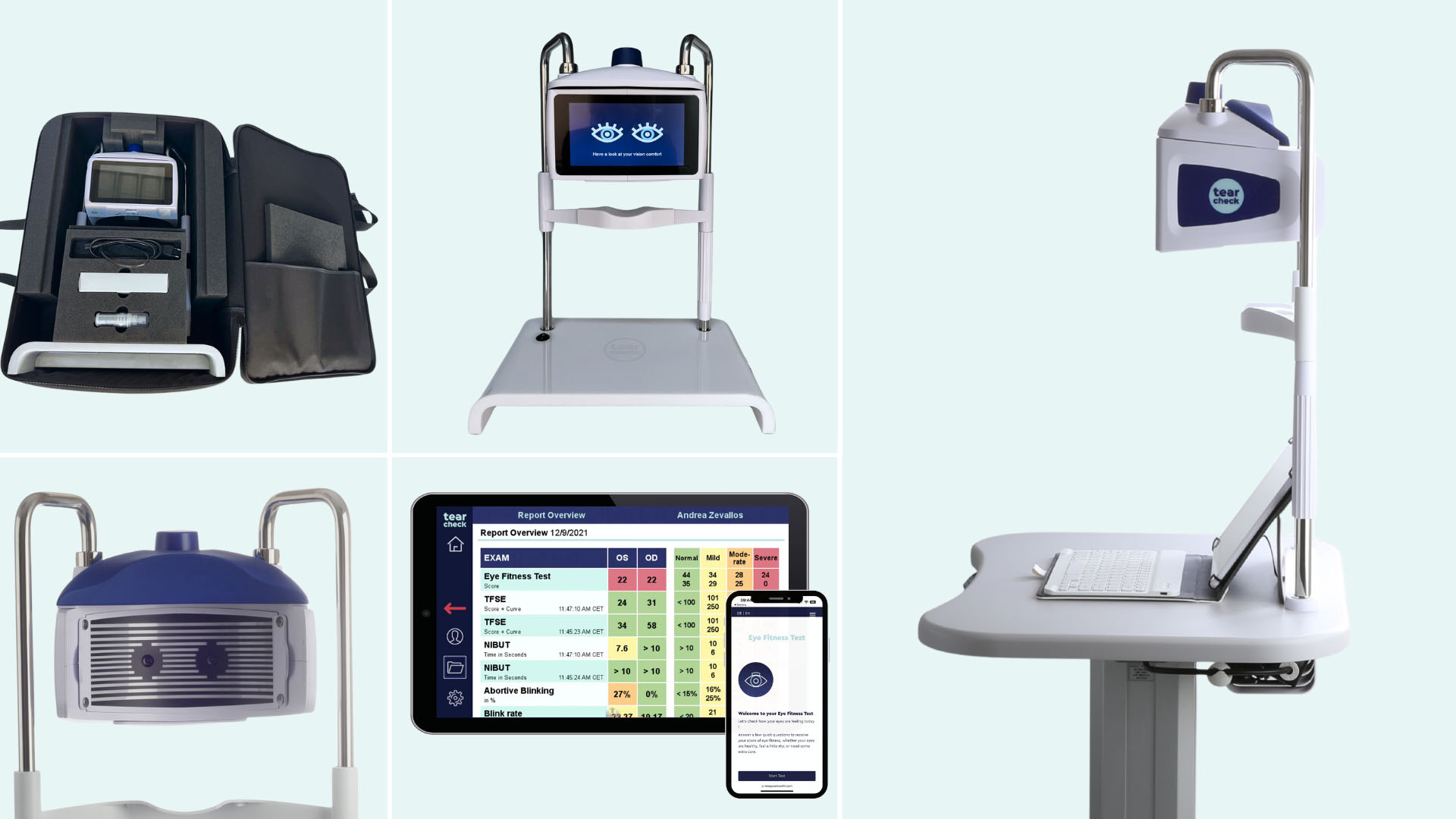
Discover tearcheck® LITE
The new tearcheck® LITE variant is available ! Experience simplified workflows within a light-weight, mobile device optimized for efficiency. Get in touch with our team to learn more !
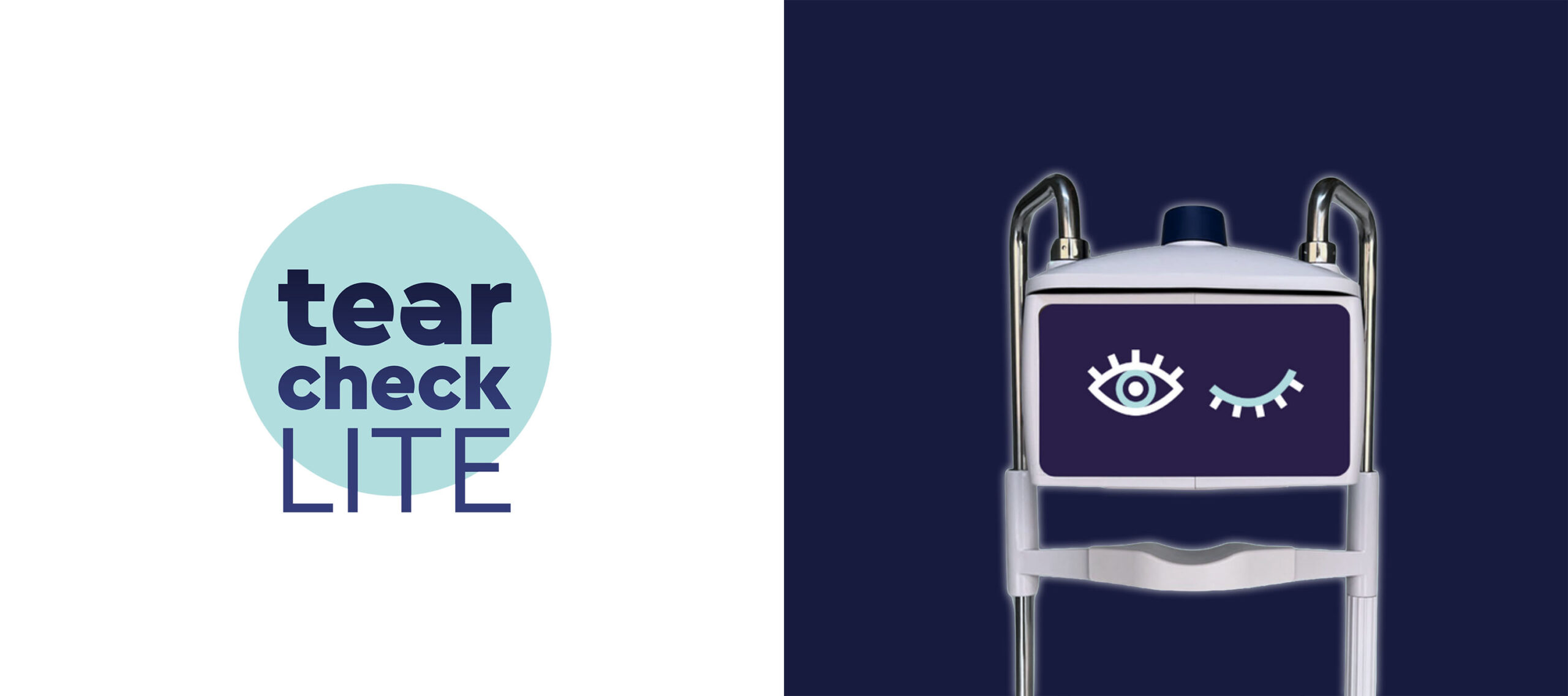
Perfect for mobility and smaller spaces

Sturdy, custom-made carry-on bag
for storage and transport
Mounted with shorter tubes,
designed to fit on refraction unit
All features are available for comprehensive
and intuitive smart dry eye screening
Downloads

User Manual
To download the user manual for your device, please log in to your connect® account. The manual will be available in your country’s language: connect®
Get in touch with our team to learn more